
Advancement of 3D Printing in Veterinary Medicine
3D printing or additive manufacturing has been around since the 1980s and is currently impacting several industries in the medical field due to its flexibility and cost-saving potential. 3D printing in veterinary medicine allows surgeons to practice a surgery before performing it on a live patient and aids in a more conducive way of communication between patients and clinicians. With the help of printers and CAD software, 3D printing DICOM files can produce a replica in a variety of materials that bear an accurate likeness to the actual anatomical structure.
Because of the development of 3D printing over the years, 3D printing in veterinary medicine has found multiple uses and applications and is growing in popularity. Additionally, 3D printing in veterinary medicine has led to significant changes in the way surgical procedures are now performed on pets. Keep reading to find out more about the impact of 3D printing in veterinary medicine today.
3D printing in Veterinary Medicine is a perfect fit for performing preoperative assessments
Years ago, one of the woes of veterinarians prior to conducting a surgery is the difficulty to fully understand a malformation until they get their hands on it which usually does not happen until they are in the process of the operation. This problem is exacerbated by the fact that our household pets are also usually relatively small. To fully prepare for a surgery, vets long needed a means to study the problem area with a physical model they can actually touch.
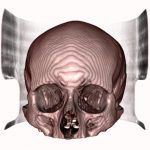
Fortunately, with the help of increasing advancement in technology, veterinarians have the capability now to 3D print CT scans of an animal’s injury or deformities that will allow them to visually assess the case and perform preoperative assessments. When vets have the chance to practice surgical procedures preceding an operation, they will be able to minimise the length of time the animal is under anaesthesia, decrease the time surgery will take from start to finish and even decrease the risk of infection. When it comes to the treatment of complex fractures, veterinarians can utilise 3D printed models to construct a detailed preoperative plan.
What’s more, researchers continue with their studies to produce full-colour models that should allow for testing new approaches that avoid contact with critical blood vessels and other tissues.
3D printed veterinary models are excellent tools for communicating with colleagues and clients
3D printing in veterinary medicine not only helps with surgical planning and visualisation but are also an advantage to the client and veterinary students.
Compared with a 3D model on a screen, an actual 3D printed model serves as an excellent tool for communicating with a client when relating to surgery or treatment a pet may undergo. Furthermore, these printed models can aid veterinarian colleagues in evaluating different approaches to solve a problem preoperatively and may help them in deciding which solution is optimal for the animal. 3D printed models can be sterilised so they can even be used during surgery as a quick reference.
Cost-effective
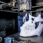
3D printing technology is a cost-effective means of making models as it has become less expensive since it was introduced in the 1980s. With a converted DICOM file and a 3D printer, a 3D model can be easily produced.
At Medimodel we aim to help clinicians establish more effective visualisation and planning prior to surgeries. We can also help with the application of scan data such as producing surgical guides, teaching aids or other uses in pre-procedure planning. Phone us on +44 (0) 117 325 8171 or reach out to us at [email protected] if you wish to discuss your case with us.

